
Figure 6. Chk1 siRNA sensitizes cells to gemcitabine.
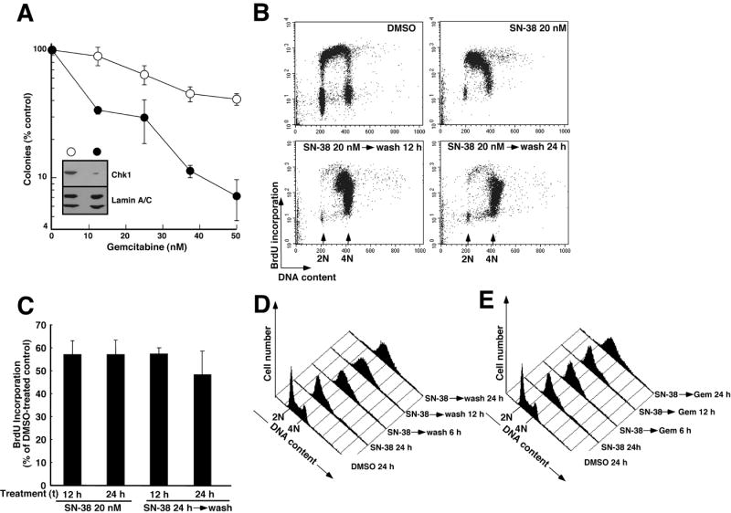
A, Ovcar-5 cells transfected twice with luciferase siRNA (open circles) or Chk1 siRNA (filled circles) were treated for 24 h with diluent (0.1% DMSO) or the indicated concentration of gemcitabine for 24 h, washed and incubated in drug-free medium for 8 d to allow colonies to form. Error bars, ± s.d. from triplicate samples. Inset in A, whole cell lysates were prepared from additional transfected cells at the time of drug treatment, subjected to SDS-PAGE and probed for Chk1. Lamins A and C served as loading controls. B, BrdU incorporation assessed during a 30 min incubation that started 12 h after addition of diluent or 20 nM SN-38. Alternatively, cells were treated for 24 h with SN-38, then washed and incubated in drug-free medium for 12 or 24 h. C, quantification of BrdU incorporation after the treatments shown in panel B. Error bars, ± S.E.M. from 4-6 independent experiments. D, histograms showing DNA content after treatment with 20 nM SN-38 for 24 h. Alternatively, cells were treated with SN-38 for 24 h, washed, and incubated in drug-free medium. Samples were harvested every 3 h after SN-38 removal. For clarity, only histograms obtained 6, 12 and 24 h after SN-38 removal are shown. E, histograms showing time-course of cell cycle progression in cells exposed to 20 nM SN-38 for 24 h, washed and treated with 10 nM gemcitabine for the indicated length of time before PI staining and analysis as indicated in panel D. Panels D and E come from the same experiment and are representative of 4 independent experiments.