
Fig. 3. Wt1-expressing epicardial cells differentiate into cardiomyocytes.
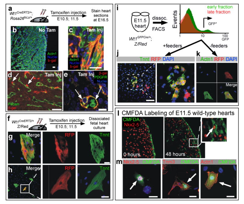
a-h. Temporal restriction of Cre labeling with Wt1CreERT2. Wt1CreERT2 did not recombine the fate mapping reporter Rosa26fsLz in the absence of tamoxifen (b). Tamoxifen treatment at E10.5 and E11.5 induced Wt1CreERT2 labeling of cardiomyocytes, as determined in both tissue sections (c) and in dissociated heart culture (g-h). Also labeled were SMCs adjacent to endothelial tubes (d-e). i-k. E11.5 Wt1GFPCre/+ heart cells actively expressing Wt1, as determined by GFP fluorescence, differentiated into cardiomyocytes. Wt1+ epicardial cells were enriched in early digestion fractions compared to late digestion fractions. GFP+ cells from early fractions were plated either with mitotically inactivated feeders (j) or without feeders (k). l-m. CMFDA dye, selectively incorporated into E11.5 epicardium, was found in CMs at 48 hours. Brief incubation of E11.5 heart explants with the dye CMFDA resulted in selective labeling of epicardium (0 hours culture). After 48 hours in explant culture, dye labeled cells were present in the myocardial wall. A subset of dye-labeled cells co-expressed the CM markers Nkx2-5, cardiac troponin I (Tnni3), and Actn1. Arrows indicate co-expression. Scale bars: 10 ⌠m.