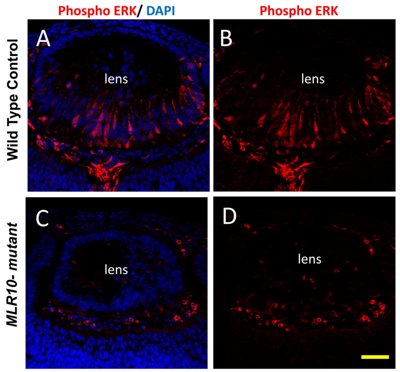
Figure 7.
Deletion of Fgfrs leads to reduction in phosphorylated Erk1/2 in the lens. Phosphorylated (active) forms of Erk1 and Erk2 were not evident in lens epithelial cells, but were readily detected in elongating primary fiber cells in control lenses (A, B). Phospho-Erk1/2 staining was dramatically reduced in the MLR10-mutant lenses (C and D) at E12.5. Nuclei were counterstained with DAPI (blue), with phosphorylated Erk1/2 staining appearing red. Scale bar = 50 μm.

