
Fig. 1. Production of VEGF, IL-6 and IL-8 as a function of tumor cell number.
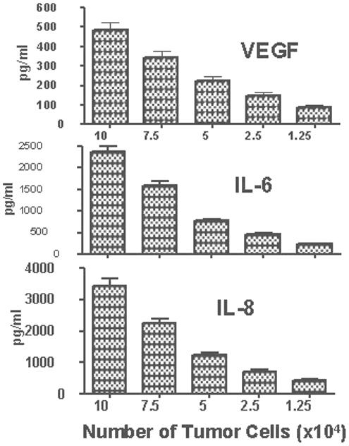
H460 tumor cells were plated onto 96-well plate at 10×104, 7.5×10⁴, 5×10⁴, 2.5×10⁴ and 1.25×10⁴cells/well. After 24 h the supernatants were collected and concentrations of VEGF, IL-6, and IL-8 were determined using multiplexed immunobeads technology. Data presented as mean concentrations (pg/ml) ± SD.