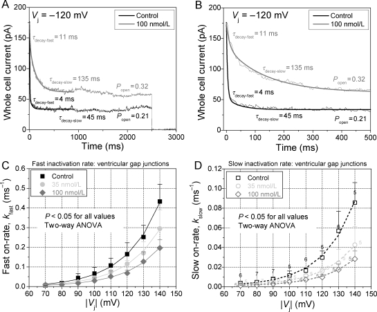
Figure 4.
Effects of rotigaptide on fast and slow ventricular Gj inactivation kinetics. (A) Biexponential fit of the decay in whole cell 2 current to obtain the fast and slow decay time constants in response to a −120 Vj step. The fast and slow inactivation on-rates were calculated under control and 100 nM rotigaptide treatments for these ventricular myocyte cell pairs of similar gj. (B) The same current traces in (A) plotted on an expanded time scale to better illustrate the exponential fit of the fast inactivation component. (C) The Vj-dependent fast inactivation rates of ventricular Gj inactivation was progressively slowed by increasing doses of rotigaptide. (D) The slow inactivation component was similarly affected by rotigaptide treatment. The number above each symbol indicates the number of experiments for the data in (C) and (D). The parameters for the fitted curves are provided in Supplementary material online, Table S2.

