
Figure 4.
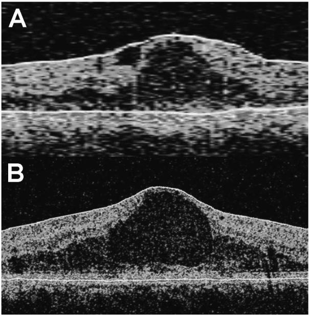
Differences in delineation of retinal layers by the Stratus OCT compared with the Cirrus HD-OCT (Carl Zeiss Meditec, Inc., Dublin, CA). Representative OCT scans obtained using the (A) Stratus OCT and (B) Cirrus HD-OCT from a study eye are shown. Segmentation lines are shown in white. The Cirrus HD-OCT segmentation algorithm identifies the thickness of the retina from the retinal pigment epithelium (RPE) to the inner limiting membrane (ILM), whereas the Stratus OCT segmentation algorithm identifies the thickness of the retina based on the distance between the ILM and junction of the outer segments (OS) and inner segments (IS) of the photoreceptors.