
Fig. 3.
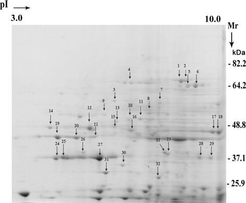
The secreted proteins ofT. vaginalis analysed by 2-D SDS-PAGE. The proteins were precipitated by TCA (Experimental procedures) for electrophoresis, and visualization of spots was by Coomassie brilliant blue staining of gels. Thirty-two proteins in the range of pH 3-10 were randomly chosen for characterization and identification by MALDI-TOF mass spectroscopy and are indicated on the gel by numbered arrows.