
Fig. 5.
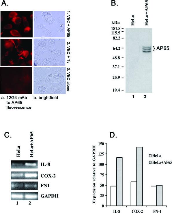
Detection of AP65 in VECs incubated with purified AP65 and live trichomonads (A and B) and induction of host gene expression in HeLa cells expressing AP65 (C and D).
A. Shows the photomicrographs of VECs from immunofluorescence using 12G4 mAb. VECs were grown on chambered culture slides and incubated with purified AP65 (panel a1) or with live T. vaginalis organisms (panel a2) for 40 min and washed well (Experimental procedures). VECs were fixed and permeabilized before incubation with mAb. The immunostained cells were observed by microscopy in oil immersion with a final magnification of 1000×.
B. Showing the nitrocellulose blot after SDS-PAGE of total proteins from control (lane 1) and transfected HeLa cells (lane 2) probed with 12G4 mAb. HeLa cells were transiently transfected with the gene encoding ap65-1. The multiple bands are the result of degradation of AP65 in the cells. No immunocrossreactive proteins were ever detected in immunoblots of control HeLa cell total proteins or control cells and cells transfected with plasmid without insert.
C. Illustrating the PCR products after agarose gel electrophoresis and EtBr staining of thetranscripts of the four genes of HeLa cells episomally expressing AP65.
Part D presents quantitative Scion image scans relative to GAPDH.