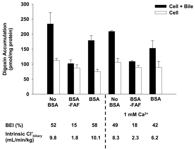
Figure 5. Effect of additional Ca2+ on the hepatobiliary disposition of digoxin in the absence or presence of BSA or BSA-FAF.
Sandwich-cultured rat hepatocytes were incubated with [3H]-digoxin (10 μM) in the absence or presence of 4% (w/v) BSA or BSA-FAF with or without 1 mM Ca2+ (calcium chloride) for 10 min following incubation in standard or Ca2+-free buffer. Filled bars represent accumulation in hepatocytes and bile canaliculi (cell + bile). Open bars represent accumulation in hepatocytes (cell). The BEI and intrinsic Cl’biliary were determined as described under Methods. Data are presented as mean ± range. n = 1 liver in duplicate.

