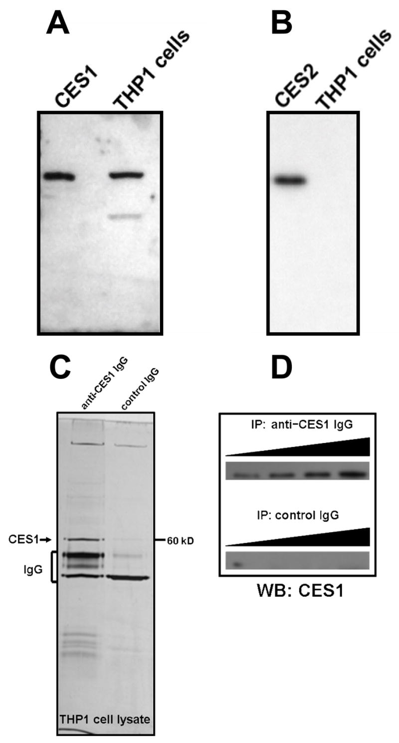
Fig. 1.

Western blot analysis of THP-1 cell lysates detects CES1 protein, but not CES2. THP-1 cell lysate (25 μg protein) was subjected to SDS-PAGE. The proteins were transferred to a PVDF membrane and probed with anti-CES1 antibody (A) or anti-CES2 antibody (B). Either 0.5 μg of recombinant CES1 (A) or 0.5 μg of recombinant CES2 (B) was included for comparison. CES1 protein can also be immunoprecipitated (IP) from soluble THP1 cell proteomes and is detected on SDS-PAGE gels following silver staining (C) or immunoblotting using anti-CES1 IgG (D). When constant amounts of THP1 cell lysate protein were titrated with increasing levels of rabbit anti-CES1 IgG and incubated, an increased yield of CES1 protein could be detected by immunoblotting in (D), while no CES1 protein was detected in incubations that used control IgG for the IP experiment (D).
