Fig. 5.

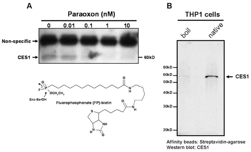
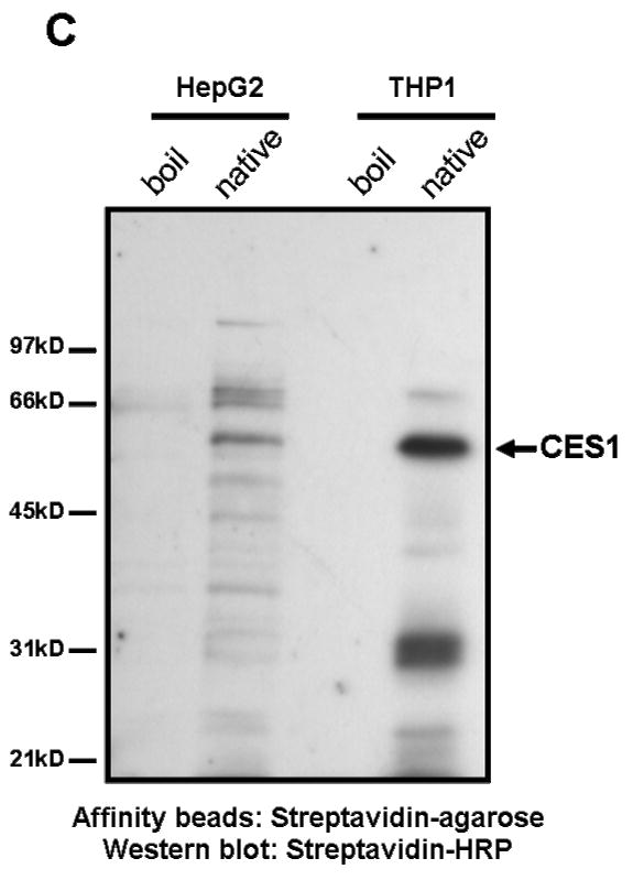
THP-1 cell lysates pre-treated with paraoxon exhibit decreased CES1-specific labeling by FP-biotin (see FP-biotin structure and chemical mechanism below blot in A). (A) Cell lysate proteins (1 mg/ml) were treated with the indicated concentrations of paraoxon for 15 min at 37°C followed by incubation with 2 μM FP-biotin for 1 h at room temperature. The lysates were subjected to SDS-PAGE, transferred to PVDF and probed with streptavidin conjugated to HRP to identify FP-biotin labeled proteins. The non-specific labeled band corresponds to a protein that reacts with FP-biotin after heat denaturation of THP-1 cell lysate proteins. The diminished band intensity of CES1 that corresponds with increasing concentrations of paraoxon indicates that CES1 is targeted by paraoxon. (B and C) Following FP-biotin-treatments of cell lysates, biotinylated proteins were enriched by incubating the lysate with streptavidin-agarose beads. Following SDS-PAGE of enriched proteins, CES1 protein was detected by rabbit anti-CES1 (B) and the biotinylated proteins detected by streptavidin-HRP (C). HepG2 cells were used to compare its suite of serine hydrolases with THP-1 cells. Boil refers to heat-denatured proteins. Native refers to non-denatured ‘active’ proteins.

