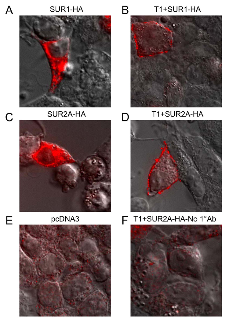
Fig. 8. Coexpression of T1 leads to the surface localization of both SUR1 and SUR2A in HEK-293 cells.
SUR1 and SUR2A were tagged with HA epitope and images from cells immunostained with an anti-HA primary and Alexa Fluor 594-conjugated secondary antibody are shown. The differential interference contrast (DIC) image is superimposed to outline the perimeters of the cells. A–D, immunostaining of SUR1 or SUR2A in the absence or presence of T1. When expressed alone, both SUR1 (A) and SUR2A (C) were localized to intracellular compartments (likely the ER). Coexpression of T1 led to the surface expression of SUR1 (B) and SUR2A (D). E–F, images from cells transfected with empty vector (pcDNA3) alone (E) and T1+SUR2A where the primary antibody was excluded in the staining procedure (F) are shown as negative controls.

