Figure 4.
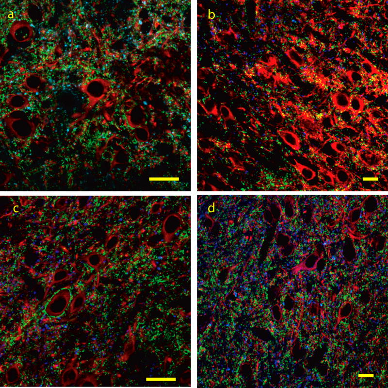
Figure 4a–d: Co-immunolabeling for VGLUT1 (blue), VGLUT2 (green) and TUJ1/MAP2 (red) in the lateral cortex (a,b) and dorsal cortex (c,d) of the inferior colliculus. In the lateral cortex both VGLUT1 and VGLUT2 labeled puncta corresponding to axo-dendritic endings are seen throughout the neuropil. In the dorsal cortex many VGLUT2 immunolabeled ending are seen surrounding the somata and proximal dendrites of medium to large neurons. Both VGLUT1 and VGLUT2 immunolabeled puncta corresponding to axo-dendritic endings are seen throughout the neuropil. Bars = 20 microns

