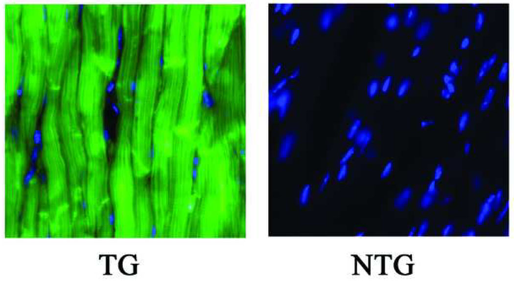
Figure 1.
Representative epifluorescence images of 10-µm sections obtained from adult hearts of an ACT-EGFP transgenic (TG) mouse and its non-transgenic (NTG) littermate. Sections were stained with Hoechst. Images for EGFP (green) and Hoechst (blue) fluorescence were merged. Imaging parameters were identical for both sections.

