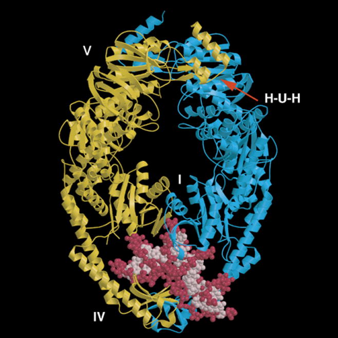
Fig. 2.

Structural model for T. aquaticus MutS bound to a mismatched DNA. The two protein monomers containing a deletion of the C-terminal 43 amino acids are shown in yellow and blue. The mismatched DNA containing a single unpaired T is shown in pink and red. Domains I and IV constitute the mismatch binding site. Two composite nucleotide binding sites reside in domain V. The H-U-H helix-u-turn-helix motif is essential for subunit dimerization.
