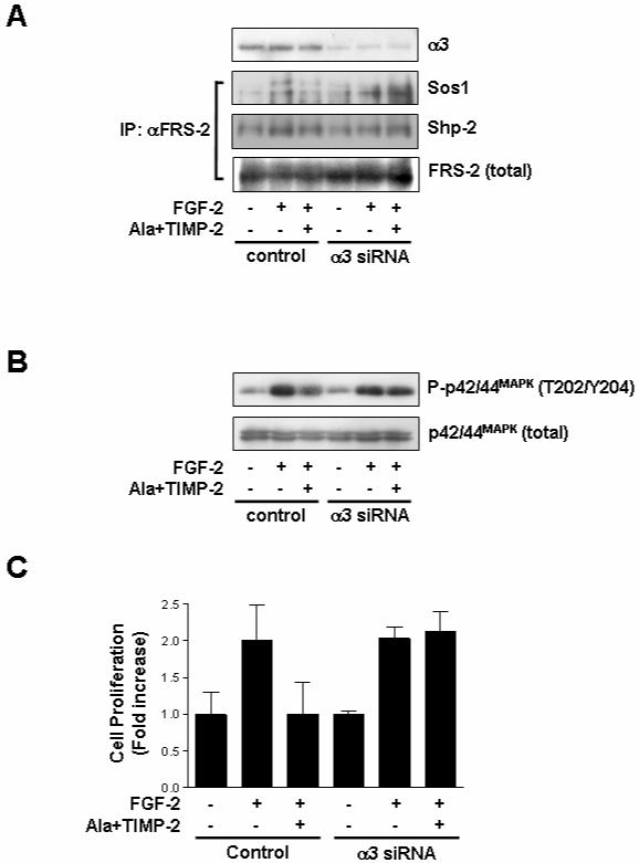
Fig. 5. Down-regulation of integrin α3 expression abrogates the suppressive effects of TIMP-2 on FGF-2-induced p42/44MAPK and cell proliferation.

Subconfluent hMVECs in gelatin-coated 100 mm dishes were transfected with integrin α3 siRNA or control siRNA as described in materials and methods. Quiescent cells were pre-treated with Ala+TIMP-2 (50 nM) for 15 min prior to FGF-2 (50 ng/ml) stimulation for 5 min (A), 15 min (B) or 24 h (C). (A) Cell lysates were Western-blotted with anti-integrin α3 (I-19) antibody. Immunoprecipitates (1mg) with anti-FRS-2 monoclonal antibodies were resolved by SDS-PAGE and Western-blotted with anti-Sos1, anti-Shp-2 or anti-FRS-2 antibodies. The results are representative of two independent experiments. (B) Cell lysates were Western-blotted with anti-phospho-p42/44MAPK or anti-p42/44MAPK antibodies. The results are representative of three independent experiments. (C) Cell proliferation results from triplicate determinations (mean ± S.D.) are presented as the fold-increase of non-treated growth.
