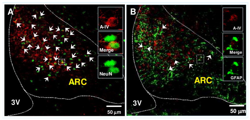
Figure 5.
Confocal image of double-immunofluorescent staining for apo A-IV (red) and cell type maker (green): NeuN (for neuronal cells, Fig 6A) or GFAP (for astrocytes, Fig. 6B) in the ARC of the hypothalamus. The colocalizations of apo A-IV with cell type marker proteins are depicted in yellow color and indicated by arrows. Solid boxes in each image are high-magnified image of the area dotted boxes. Sections are representative of 4 animals in which staining was examined. 3V: Third ventricle.

