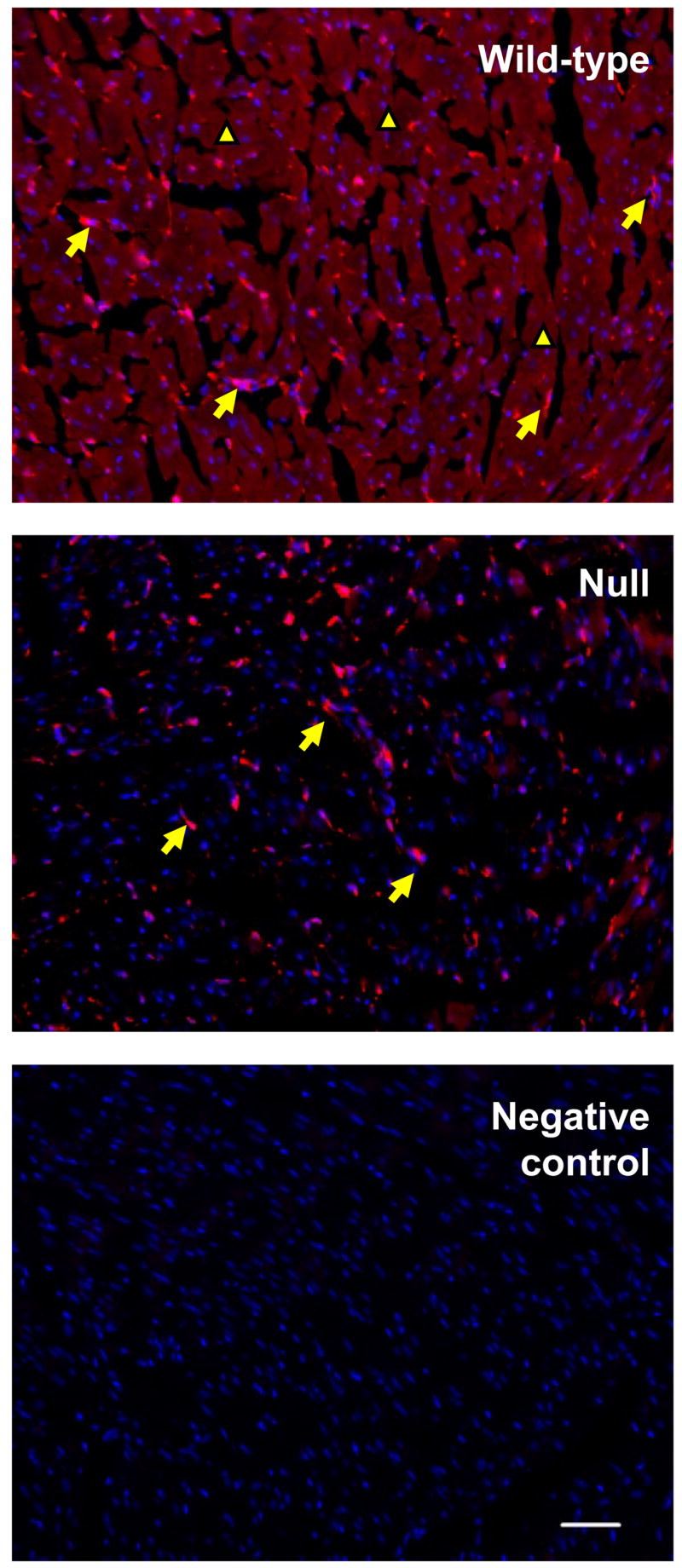
Fig. 2.

Immunofluorescent detection of CPR in the wild-type and cardiomyocyte-Cpr-null hearts. Frozen sections (10-μm thick) from 2-month-old male mice were incubated with a rabbit anti-CPR antibody. CPR expression was visualized by a chicken anti-rabbit secondary antibody conjugated with Alexa 594 (red fluorescence). The sections were counterstained with DAPI (blue fluorescence). The anti-CPR antibody was omitted in the negative control section. CPR immunostaining was found in cardiomyocytes (small arrowheads) as well as other cell types, including cells that appear to be cardiac fibroblasts (large arrowheads). Bar = 50 μm. The results shown are typical of sections from seven mice for each strain.
