
Fig. 6. Notch1 signaling augments Wnt signaling in the otic placode.
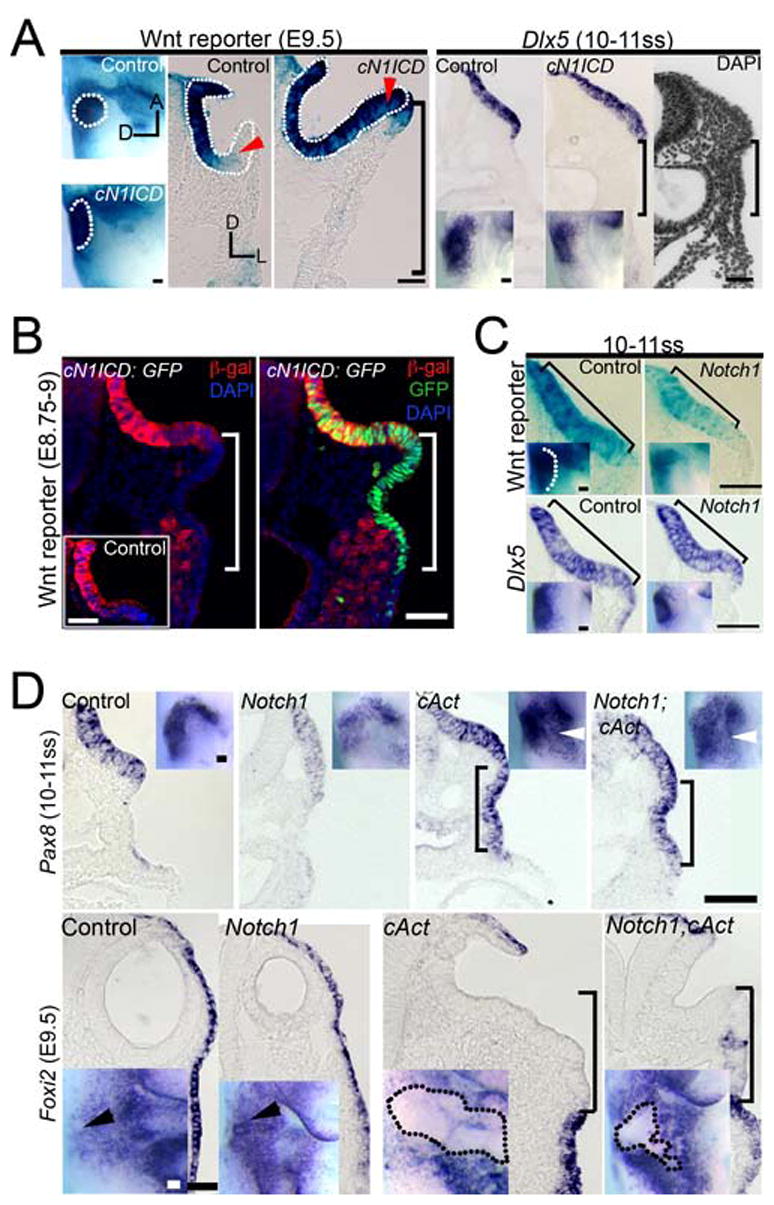
(A) Wnt reporter and Dlx5 mRNA expression is increased in conditionally over-expressing Notch-ICD (cN1ICD) embryos in whole-mount (top row) and corresponding mid-placode transverse sections (bottom row). Dotted outline: otic cup. Red arrowhead: medio-lateral otic region. (B) Wnt reporter expression in transverse sections of control and cN1ICD; Wnt reporter embryos; Wnt reporter mice co-immunostained with anti-β-galactosidase (β-gal; red) and anti-GFP (green) antibodies. Note that only the medial part of the expanded placode expresses the Wnt reporter. The inset shows anti-β-galactosidase staining in a normal Wnt reporter mouse. (A–B) Bracket: ectopic lateral placode region is negative for Wnt reporter and Dlx5. (C) Wnt reporter and Dlx5 expression is diminished in Notch1 mutants relative to controls. Brackets: otic placode. (D) A comparison of Pax8 and Foxi2 expression in Notch1 mutant, cAct and Notch1; cAct double mutant littermates. (A, C, D) Arrowhead: otic expression. Scale: 50 μm.