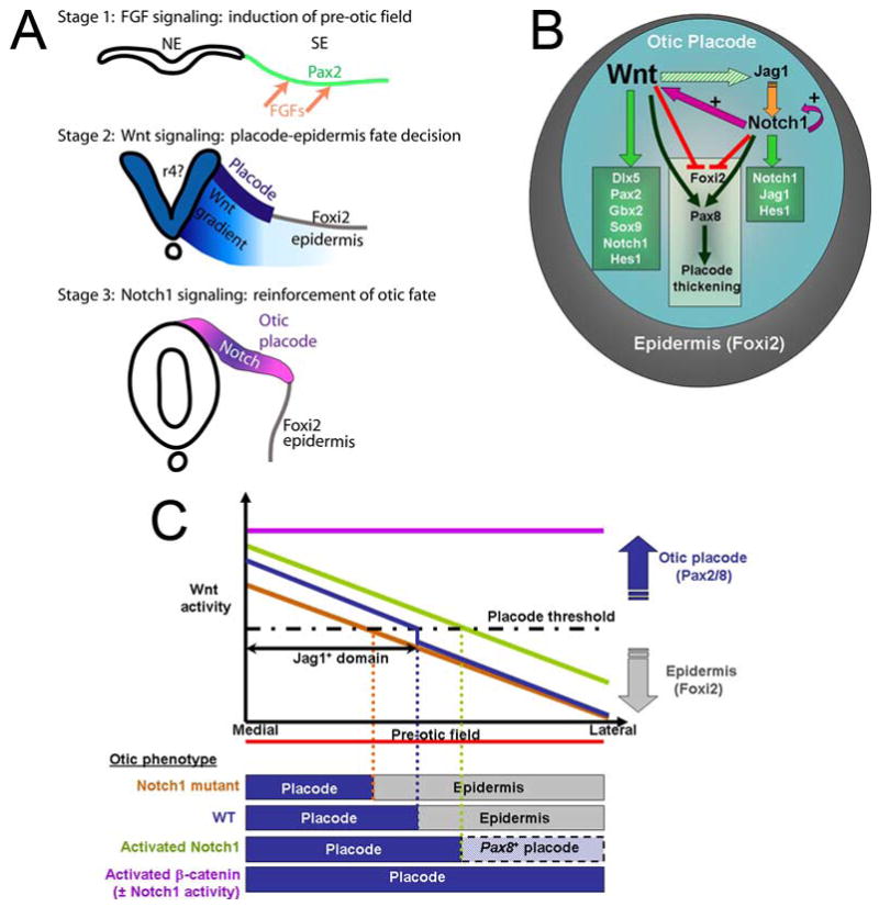
Fig. 7. Model of how Wnt and Notch pathways interact to regulate the size of the otic placode.

(A) The generation of the otic placode can be divided into three stages. Pax2 in the pre-otic field is induced by FGFs (arrows). A gradient of Wnts (light blue) determines the size of the otic field; above a certain threshold, Wnts drive cells towards an otic fate (dark blue) and below the threshold, cranial epidermis is formed (Foxi2) (Ohyama and Groves, 2003). Notch1 signaling is superimposed on the Wnt gradient (pink-blue) and acts to augment otic fate imposed by Wnts. NE; Neuroectoderm, SE; Surface ectoderm (this paper). (B) The Wnt pathway is the primary signal (denoted by bold lettering) that controls otic fate (blue region) by positively regulating (green arrows) the expression of Dlx5, Sox9, Gbx2, Pax2/8 and components of the Notch1 pathway such as Notch1 and Hes1 (Figs. 1 and 2). Jag1 expression is initiated by Wnts (dashed green arrow) (Fig. 2). Notch1 acts to: (1) augment Wnt and Notch1 activity within otic cells (pink arrow; plus sign) and (2) co-operate with Wnt to negatively regulate Foxi2 (red) and positively regulate Pax8 (dark green) and maintain a thickened otic placode. (C) A model summarizing the various otic placode phenotypes observed in this study. A gradient of Wnt activity emanating from the midline is established across the medio-lateral axis of the pre-otic field. Cells exposed to a certain threshold of Wnt signals express Jag1 and differentiate as otic placode (blue). Below this threshold, cells differentiate as epidermis (grey). Jag1-Notch1 signaling augments Wnt signals in the medial region of the otic placode, whereas more lateral regions are not exposed to Notch1 signals and Wnt signaling is not augmented. In the absence of Notch1 (yellow line), the gradient of Wnt signaling becomes weaker, resulting in a smaller placode and more epidermis. When Notch1 is activated in the pre-otic field (green line), the Wnt gradient is augmented further. Some Wnt-dependent markers (Dlx5) are expressed only in the expanded Wnt domain, whereas markers such as Pax8 are expressed throughout the pre-otic field (marked as lateral placode). When β-catenin is activated in the entire pre-otic field (purple line), all cells differentiate as otic placode (Ohyama et al., 2006).
