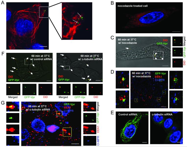
Figure 4.
Microtubule-associated transport of viruses. (A) Colocalization of GFP-Vpr-labeled lentiviruses with microtubule networks 1 h after infection. Microtubules were immunostained with the monoclonal antibody to α-tubulin (red). The boxed region is enlarged in right panel. Arrows indicate viruses on the microtubules. (B) Microtubule staining in a nocodazole-treated cell. Cells were preincubated with nocodazole at 37°C for 30 min to disrupt microtubules, then immunostained with the monoclonal antibody to α-tubulin (red). (C) The fixed image of GFP-Vpr/DiD-labeled viral particles in a nocodazole-treated cell (see Materials and Methods). Arrows indicate the viral particles fused to endosomes. (D) Localization of GFP-Vpr-labeled viral particles (FUW-GFPVpr/αCD20+SINmu) with the two endosomal markers after 60 min of incubation in a nocodazole pre-treated cell. (E) Microtubule staining of siRNA-treated cell. 293T/CD20 cells were transfected with α-tubulin-specific siRNA- or control siRNA. After 72 h, transfected cells were seeded and immunostained with anti- α-tubulin antibody (green). (F) The virus fusion for siRNA-treated cells. α-tubulin siRNA or control siRNA-treated cells were incubated with GFP-Vpr/DiD-labeled viruses at 37°C for 60 min and then fixed. Yellow particles denoted by arrows indicate the virus particles fused to endosomes. (G) Localization of GFP-Vpr-labeled viral particles with the two endosomal markers after 60 min of incubation with α-tubulin siRNA-treated cells. The boxed regions are enlarged and shown in separated panels. Scale bar represents 5 μm.

