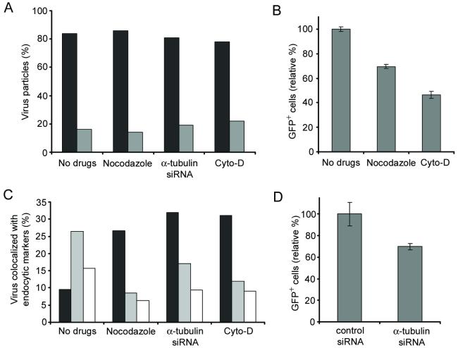
Figure 5.
The effects of inhibitory drugs or siRNA treatment on viral fusion, infection, and endosome maturation. (A) GFP-Vpr/DiD-labeled viruses were incubated with drug- or siRNA-treated cells at 37°C for 60 min, and then fixed. The viral particles with the fusion signal (black) or without the fusion signal (gray) were quantified. The viral particles both GFP-Vpr+ and DiD+ were considered to be fused with endosomes, while particles that were only GFP-Vpr+ were considered to be unfused virus. For quantification, 60 viral particles were examined for no drug-treatment, 64 particles for nocodazole treatment, 72 particles for α-tubulin siRNA treatment, and 68 particles for cyto-D treatment. The results were collected from three independent experiments. (B) The role of microtubules and actin filaments in the virus infection. 293T/CD20 cells which were preincubated with nocodazole or cytochalasin-D (cyto-D) were transduced with 2 ml of fresh unconcentrated FUGW/αCD20+SINmu virus. The resulting GFP expression was analyzed by FACS. (C) Quantification of GFP-Vpr-labeled viruses colocalized with EEA1+ (black), CI-MPR+ (white), or both EEA1+ and CI-MPR+ (gray) endosomes at 60 min of incubation in drug- or siRNA-treated cells. (D) The effect of α-tubulin knockdown on virus infection. Control siRNA or α-tubulin siRNA transfected cells were transduced with 2 ml of fresh unconcentrated FUGW/αCD20+SINmu virus. The percentage of GFP+ cells was analyzed by FACS.

