
Fig. 1.
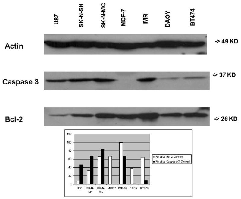
Representative Western Blot. Lysates of cultured SK-N-SH, SK-N-MC, and IMR-32 neuroblastoma cells, BT474 and MCF-7 breast cancer cells, U87 glioma cells, and DAOY medulloblastoma cells were applied to an SDS-polyacrylamide gel and transferred to a membrane for Western blotting as described in Materials and Methods. The same blot was stained with antibodies to Bcl-2, caspase-3, and β-actin, respectively. Western blot bands obtained for each cell lysate were subjected to optical densitometry and the values for Bcl-2 and caspase-3, respectively, were normalized to the value for β-actin on the same blot. Results are expressed as relative optical densities.