
Fig. 2.
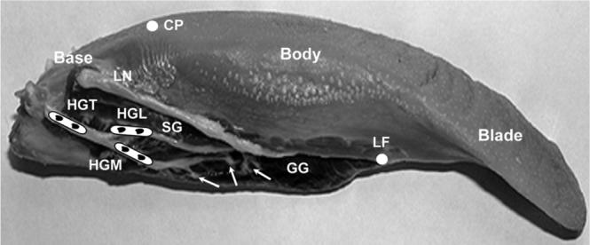
Scheme for the hypoglossal nerve and the extrinsic muscles stimulation. White solid circles indicate the anatomical landmarks for dividing the tongue into three segments: Blade: tongue tip to lingual frenum (LF); Body: lingual frenum to circumvallate papillae (CP); Base: papillae to tongue posterior border. HGT, hypoglossal trunk; HGM, medial branch; HGL, lateral branch; GG, genioglossus; SG, styloglossus; LN, lingual nerve. Arrows indicate secondary nerve branches of the HGM to GG. Black dots indicate the sites of stimulation electrodes.