
Fig. 3.
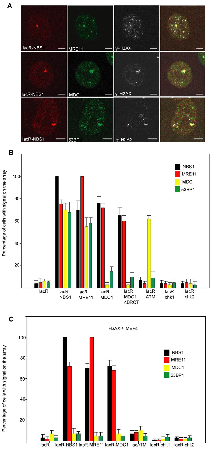
Cross-recruitment of repair factors. (A) Immunofluorescence microscopy on NIH 2/4 cells transiently transfected for 24h with lacR-NBS1 (red) with the indicated antibodies (green). Scale Bar = 2µm. (B) Quantitation of repair factor accumulation on the array in NIH 2/4 cells after immobilization of the indicated repair factors. Values represent averages ± S.D (n = 40) from 3 independent experiments. The antibody used to detect MDC1 recognizes only the mouse isoform and not human MDC1 present in the LacR fusion protein. (C) Quantitation of repair factor accumulation on the array on H2AX−/− MEFs containing the lacO array after immobilization of the indicated repair factors. Values represent averages ± S.D (n = 50) from 3 independent experiments.