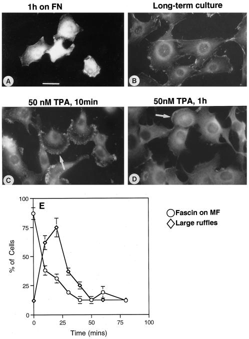
Figure 1.
Fibronectin adhesion and TPA treatment alter fascin localization in C2C12 cells. C2C12 cells were stained for fascin after 1 h of adhesion on fibronectin in serum-free medium (A), in long-term culture 1 h after addition of DMSO solvent control (B), after 50 nM TPA for 10 min (C), and after 50 nM TPA for 1 h (D). An example of a large ruffle is arrowed in C, and the residue of a ruffle is arrowed in D. Bar, 10 μm. (E) The percentage of cells with microfilament-associated fascin (○) or large ruffles (⋄) was quantitated with time of TPA treatment. Each point is the mean ± SEM of four independent experiments.

