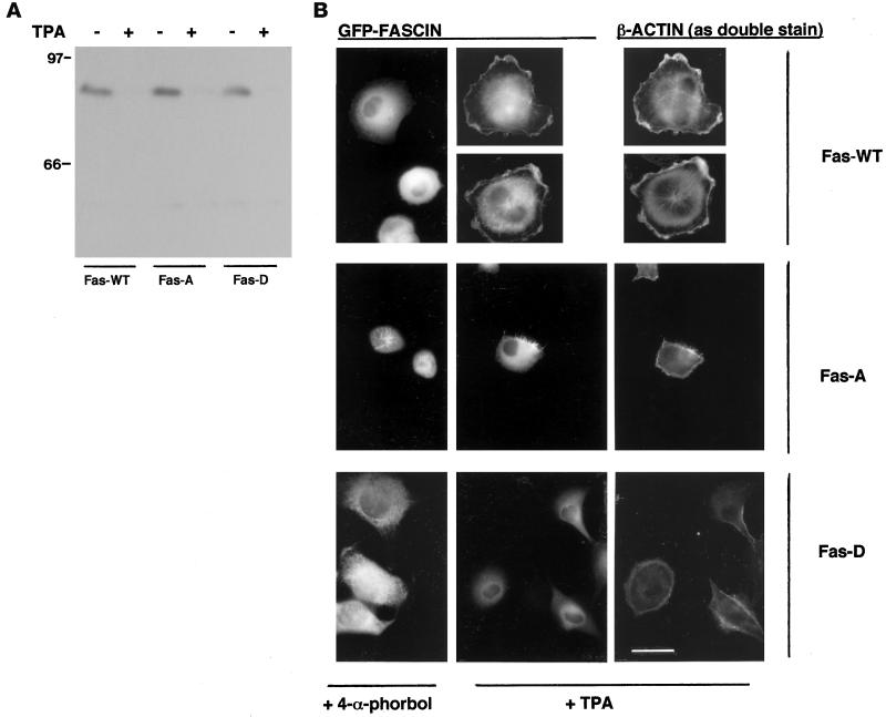
Figure 11.
Requirements for PKCα activity and fascin phosphorylation at serine 39 in matrix-dependent fascin localization in LLC-PK1 cells. (A) Western blot of whole cell extracts of LLC-PK1 cell lines expressing GFP-fascin (Fas-WT), GFP-S39A fascin (Fas-A), or GFP-S39D fascin (Fas-D) prepared after 24 h of treatment with 100 nM 4-α-phorbol (TPA −) or 100 nM TPA (TPA +). The blot was probed with antiserum to PKCα. (B) Comparison of GFP-fascin localization in cells treated with 100 nM 4-α-phorbol or TPA for 24 h after 1 h of adhesion to fibronectin under serum-free conditions. TPA-treated cells are shown double stained for β-actin. Bar, 5 μm.

