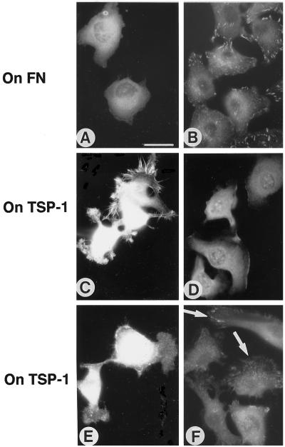
Figure 3.
Effect of PKC activation on microspikes and focal contacts in matrix-adherent C2C12 cells. (A and B) Cells adherent for 1 h on fibronectin in the presence of 50 nM TPA. (C and E) Control C2C12 cells adherent for 1 h on TSP-1. (D and F) Cells adherent on TSP-1 for 1 h in the presence of 50 nM TPA. (A, C, and D) Fascin stain. (B, E, and F) Vinculin stain. Arrows in F indicate localization of vinculin in focal contacts. Bar, 10 μm.

