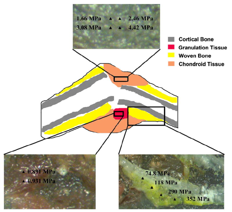
Fig. 2.

Schematic of a representative callus section depicting the location of the different tissue types with respect to the fracture gap and pre-existing bone. Indentation moduli at multiple positions throughout regions of each tissue type are shown in the insets.
