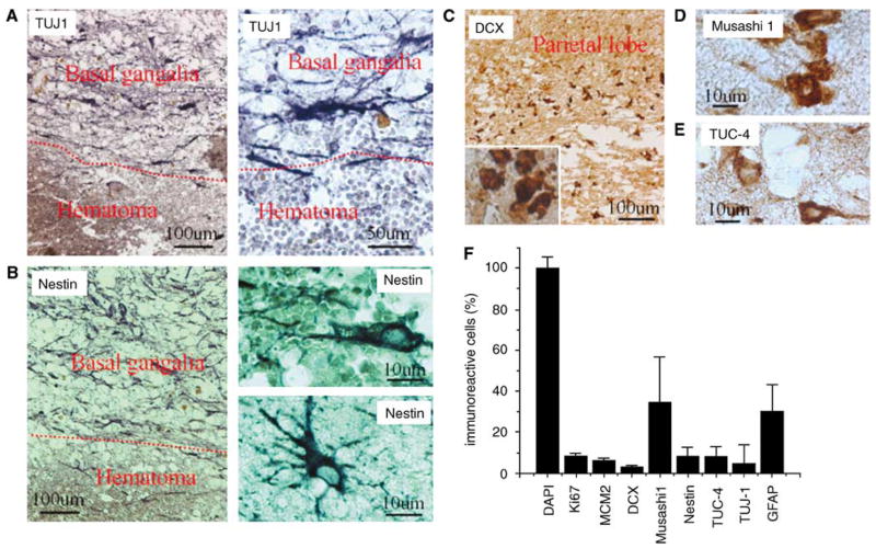
Figure 1.
Expression of NSC marker proteins in the perihematomal region after ICH in adult human brain. (A) TUJ-1 shown at low (left) and high (right) magnification. (B) Nestin shown at low (left) and high (right) magnification, both adjacent to (top right) and at a distance from (bottom right) the hematoma. (C) DCX at low and high (inset) magnification. (D) Musashi1. (E) TUC-4. (F) The number of NSC-positive cells and proliferative cells in the perihematomal region after ICH.

