
Figure 3.
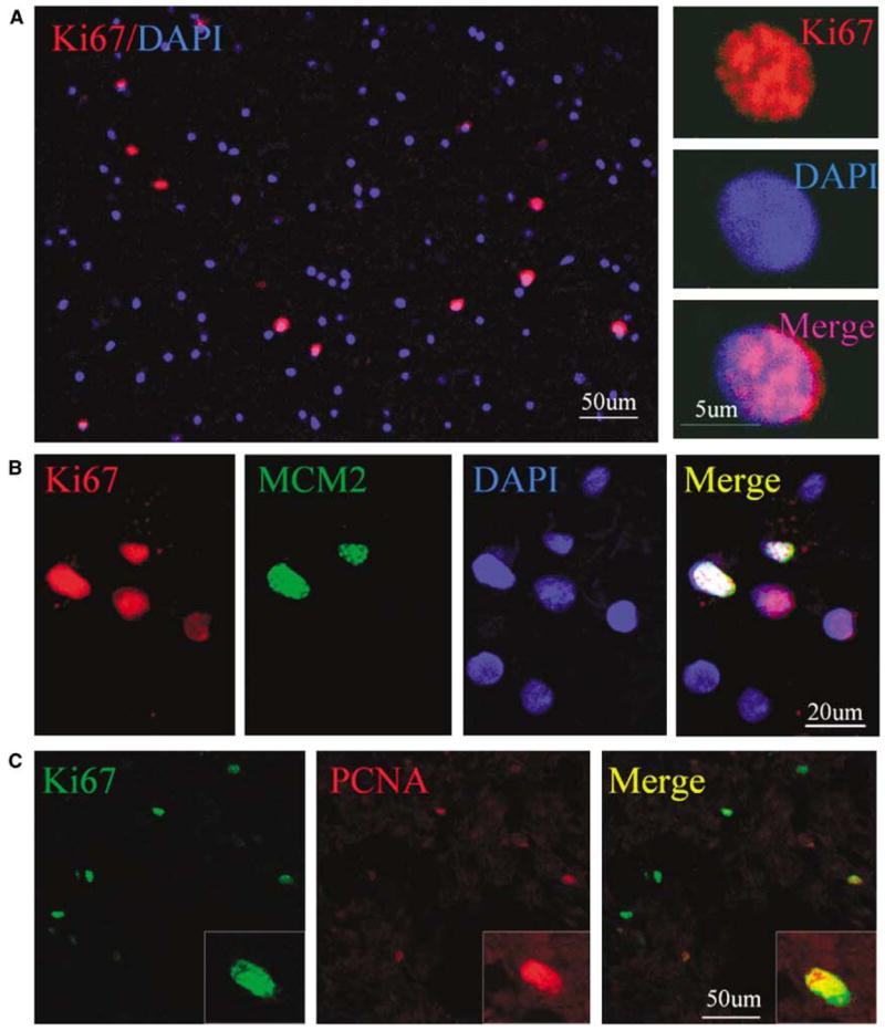
Presence of proliferative cells in perihematomal region after ICH. (A) Ki67-positive cells (red) are shown in relation to all cells (DAPI-stained nuclei, blue) at low (left) and high (right) magnification. (B) Ki67 (red) is colocalized with minichromosome maintenance 2 (green) in the nucleus (DAPI staining, blue) of cells adjacent to the hematoma. (C) Ki67 (green) is colocalized with proliferating cell nuclear antigen (red) in the nucleus of a perihematomal cell (seen at far right in low-magnification panels).