
Figure 5.
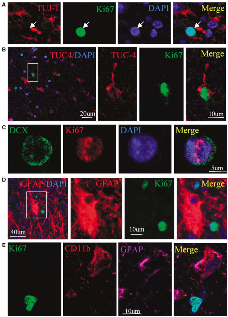
Coexpression of neuronal lineage and cell proliferation markers in perihematomal regions after ICH. (A) A section stained for TUJ-1 (red cytoplasm) and Ki67 (green nucleus) shows that TUJ-1-positive cells express Ki67. (B) A TUC-4-positive (red cytoplasm) cell also expresses Ki67 (green nucleus). (C) DCX (green cytoplasm) is colocalized with the cell proliferative marker Ki67 (red nucleus). (D) GFAP-positive (red cytoplasm) cells do not colocalize with Ki67 (green nucleus). (E) CD11b-positive (red cytoplasm) cell expresses GFAP (purple) but not Ki67 (green). Nuclei in all panels are counterstained with DAPI (blue).