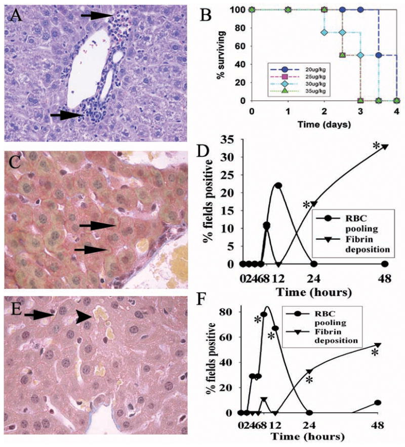
Figure 2.

Hepatic injury and lethality in a mouse model of ricin intoxication. A. H&E stain of peri-portal liver 48 hours following ricin challenge, showing nests of leukocytes (arrows) adjacent to portal tracts. B. Lethality dose-response after ricin administration i.p. at the doses indicated, with 10 mice per group. C. Fibrin in the peri-portal area of liver 48 hours after ricin challenge (40 μg/kg). Deposition of fibrin (arrows) occurred between hepatocytes and was detectable by MSB staining (see Materials and Methods). D. Time course of fibrin deposition in the peri-portal liver after ricin challenge. This is shown as the percent of high-powered (400×) microscopic fields showing fibrin deposits around hepatocytes. E&F, same as C&D, except that the centro-lobular liver was studied. RBC pooling (arrowhead in panel E) refers to aggregates of red blood cells two or more times larger than present in normal liver sinusoids, and scored as the percent of high-powered fields positive for the finding. For D & F, findings are representative of two independent studies, with a total of 4 mice per time point. *, p< 0.04, comparing number of fields showing fibrin deposition in centro-lobular and peri-portal areas at 24 and 48 hours with that at 0–4 hours. **, p<0.05, comparing number of fields showing RBC congestion at 0 and at 8 hours. Magnification: A, C, and E, 400×.
