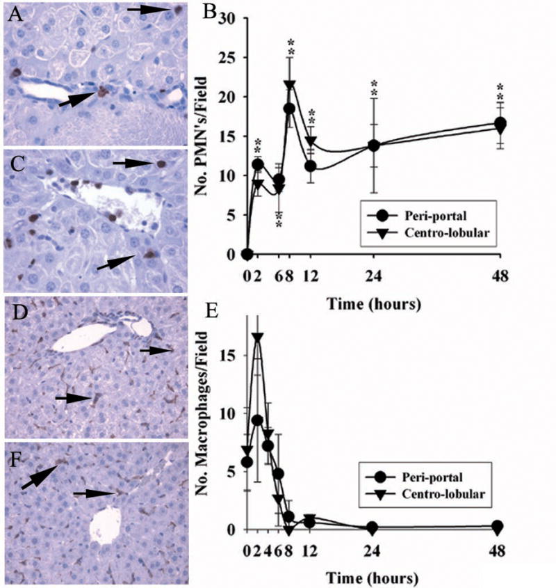
Figure 3.

Migration of leukocytes to liver after ricin challenge in vivo. A & C. Immuno-reactive neutrophils in peri-portal ( panel A) and centro-lobular ( panel C) areas of liver 48 hours after toxin administration. B. Quantification of neutrophils as number of 7/4+ immuno-reactive cells per high-powered field 0–48 hours after toxin was given. D & F. Immuno-reactive macrophages in peri-portal (panel D) and centro-lobular (panel F) hepatic areas 2 hours after toxin challenge. E. Number of F4/80+ immuno-reactive cells per HPF of liver 0–48 hours after administration of toxin. Repeat studies showed loss of stained cells occurred over 8–12 hours. Findings are representative of two independent studies with a total of 4 mice per time point. Magnification: 400× (A, C); 200× (D, E). *, p < 0.01, number of leukocytes per HPF at the designated hours compared with that at 0 hours. **, p < 0.02, number of macrophages per HPF at 0 hours compared with that at 2 hours in the peri-portal area.
