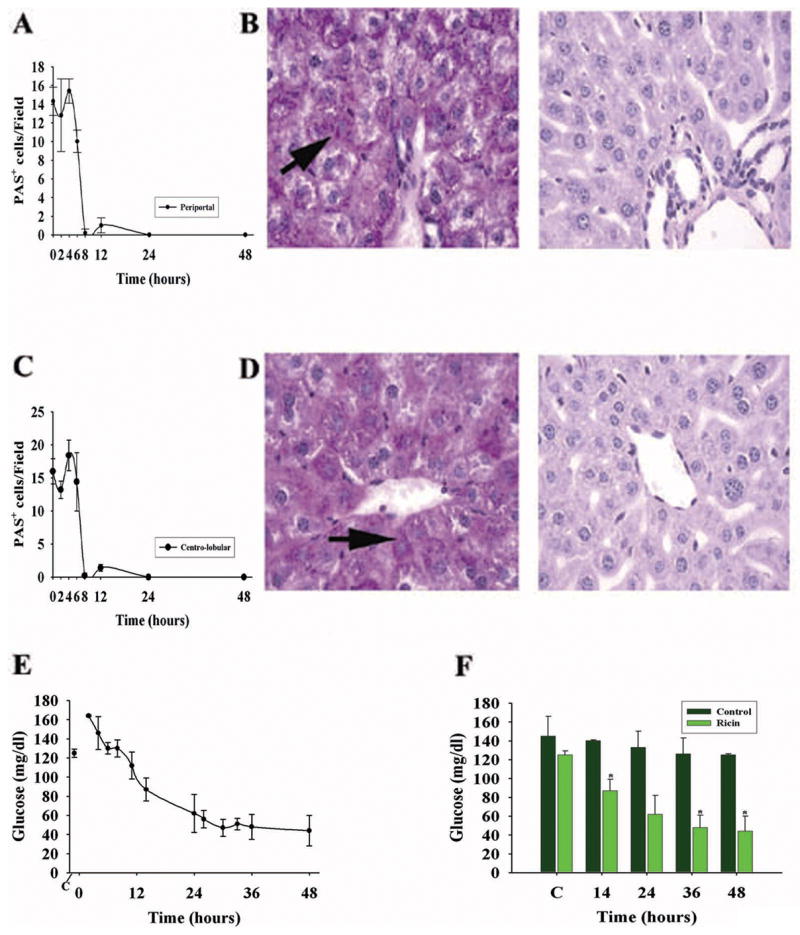
Figure 5.

Hepatic glycogen and hypoglycemia following ricin challenge (40 μg/kg) in the C57BL/6 mouse. A. Time course for the presence of peri-portal glycogen-containing hepatocytes, displayed as the number per high power field, detected as periodic acid Schiff (PAS+) -positive cells. B. PAS staining of peri-portal hepatic parenchyma at 0 and 8 hours (B, left and right panels, respectively) after ricin exposure. Arrows, individual PAS+ hepatocytes. C. Number of PAS+ cells per HPF in the centro-lobular liver from 0 to 48 hours following ricin. D. Examples of PAS+ hepatocytes at 0 and 8 hours (D, left and right panels, respectively) following toxin. For A & C, results are representative of two independent studies, each with 2 mice per time point (total of 4 mice per time point, or 32 mice total). Complete disappearance of glycogen took up to 12 hours in repeat studies. PAS+ cell number at 8–48 hours was significantly different from that at 0 hours (p <0.001). E. Blood glucose in mice following ricin challenge, as determined by glucometer on tail vein blood. F. Comparison of blood glucose levels in saline (control)- and ricin-challenged mice at selected times before and after ricin challenge. *, p < 0.02, comparing ricin-treated versus vehicle-treated mice at the indicated time points (14, 36, and 48 hours). C, blood glucose prior to challenge with ricin or vehicle. Magnification: B, D (400×).
