Figure 3.
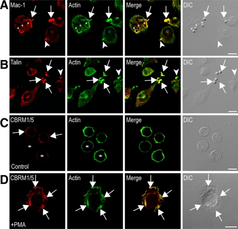
Mac-1 and talin localize to actin-rich membrane ruffles engaged in Mac-1–mediated phagocytosis. RAW264.7 macrophages stimulated with 10 μg/ml LPS were exposed to C3bi-sRBCs and immunostained for (A) Mac-1 (red) and actin (green) or (B) talin (red) and actin (green) as described in Materials and Methods and imaged using confocal microscopy. Merged confocal and corresponding DIC images are also shown. Arrows in (A) indicate clustering of Mac-1 into actin-rich membrane ruffles, adjacent to the site of particle attachment. Circular Mac-1 and actin colocalization was also visualized around recently internalized particles (asterisks). Arrows in B illustrate prominent talin and actin colocalization at membrane ruffles surrounding bound C3bi-sRBCs. Arrowheads indicate Mac-1/talin staining enrichment in membrane ruffles not containing C3bi particles. Differentiated U937 cells stimulated without (C) or with 150 nM PMA (15 min; D) were immunostained for activation-specific Mac-1 epitope using CBRM1/5 (red) and actin (green), as described in Materials and Methods, and imaged using confocal microscopy. Merged confocal and corresponding DIC images are also shown. (C) Asterisks indicates cells with negligible CBRM1/5 antibody staining; arrows indicate punctate staining at random sites on the cell periphery. (D) Arrows indicate the high-affinity integrin enrichment in actin-containing membrane ruffles in PMA-stimulated cells. Scale bars, 10 μm.

