Figure 2.
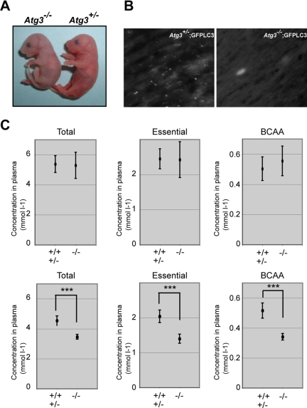
Phenotypes of Atg3-deficient mice. (A) Morphology of Atg3+/- and Atg3−/− mice. (B) Deficiency of LC3-positive dots in Atg3−/− heart. Atg3+/− and Atg3−/− mice expressing GFP-LC3 were delivered by Cesarean section and analyzed by florescence microscopy. Representative results obtained from each neonatal heart at 3 h after Cesarean delivery. Bar; 50 μm. (C) The concentrations of total amino acids, essential amino acids, and BCAA in sera of mice of the indicated genotypes. Atg3+/+, Atg3+/− (n = 5) and Atg3−/− mice (n = 4) were dissected immediately (top) or at 10 h (bottom) after delivery and the plasma concentrations of amino acids were measured. Data are mean ± SD. ***p < 0.001 by Student's t test.

