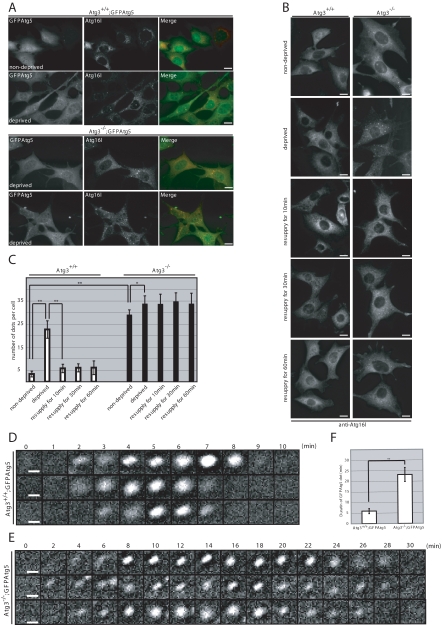
Figure 4.
Accumulation of Atg16L-positive structures in Atg3-deficient MEFs. (A) Immunofluorescence analysis of Atg16L in GFPAtg5-introduced immortalized MEFs. Each genotype MEFs were cultured in nutrient-rich (nondeprived) or Hanks' solution (deprived). The cells were fixed and then immunostained with anti-Atg16L. Bars; 10 μm. (B) Immunofluorescence analysis of Atg16L in MEFs. MEFs isolated from Atg3+/+ and Atg3−/− were cultured in nutrient-rich (nondeprived) or Hanks' solution (deprived). After deprivation of nutrients, the cells were resupplied with nutrients for 10, 30, or 60 min. The cells were fixed and then immunostained with anti-Atg16L. Bars; 10 μm. (C) The number of Atg16L-positive dots in MEF (n = 20) was determined in each genotype. Data are mean ± SD. *p < 0.05 and **p < 0.01 by Student's t test. (D and E) Time-lapse observation of starvation-induced GFPAtg5 dots. Wild-type (D) and Atg3-deficient (E) MEFs harboring GFPAtg5 were cultured in Hanks' solution for 1 h and directly observed by time-lapse video microscopy. Bars, 1 μm. (F) Duration of existence of GFPAtg5 dots in wild-type and Atg3-deficient MEFs. The time of sustained presence of GFPAtg5 dots in wild-type (n = 9) and mutant (n = 10) was measured. Data are mean ± SD. **p < 0.01 by Student's t test.

