
Figure 1.
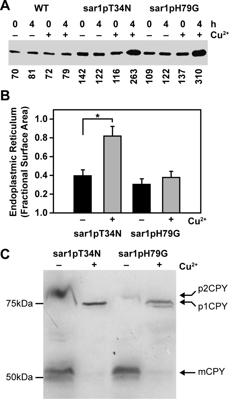
Effects of sar1pT34N and sar1pH79G on the morphology of the endoplasmic reticulum and Golgi apparatus. (A) WT, WDY64, and WDY65 cells were grown in YNM and then switched to YND (± CuSO4) for 0 and 4 h. Equal amounts of SDS solubilized cells (OD600 = 0.3) were then evaluated by Western blotting using rabbit anti-Sar1p antibodies (gift from Dr. Ben Glick). The relative amounts of Sar1p were quantified using ImageJ software. The data represent one of three trials. (B) WDY64 and WDY65 cells were adapted from YNM to YND (± CuSO4) for 4 h. The cells were processed for electron microscopy, and images were taken on a JEOL 100CX microscope. The surface area of the endoplasmic reticulum was quantified using morphometric protocols and ImageJ software. The data represent mean ± SE (n = 12–14 cells). The statistical differences relative to untreated control were determined by Student's t test (*p = 0.001). (C) WDY64 and WDY65 cells were grown in minimal YNDH medium and then switched to nitrogen-starvation SD(-N) medium (± CuSO4) for 8 h. The cells were solubilized and precursor (p1CPY and p2CPY) and mature (mCPY) forms of carboxypeptidase Y, identified by their molecular sizes on Western blots.