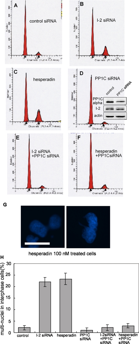
Figure 6.
Mitotic defects produced by I-2 knockdown are mimicked by Aurora B inhibition and rescued by partial PP1C knockdown. (A–F) ARPE-19 cells were transfected for 72 h with siRNA against luciferase, as control (A), I-2 (B), PP1Cα (D), or both I-2 and PP1Cα (E). For hesperadin treatment, 100 nM hesperadin was added to the medium 42 h after transfection with control siRNA (C) or siRNA against PP1Cα (F), and cells were incubated for another 30 h. The DNA content of cells was measured by flow cytometry, with propidium iodide staining. Profiles are representative of three independent experiments. Knockdown of PP1Cα was measured by blotting with anti-PP1Cα antibody, by using actin as loading control (inset in D). (G) Hoechst 33342 staining showed cells with multiple nuclei after 100 nM hesperadin treatment. Bar, 25 μm. (H) Percentage of cells with multiple nuclei after treatment with siRNA or hesperadin (500 total/group), plotted as mean ± SD from three independent experiments.

