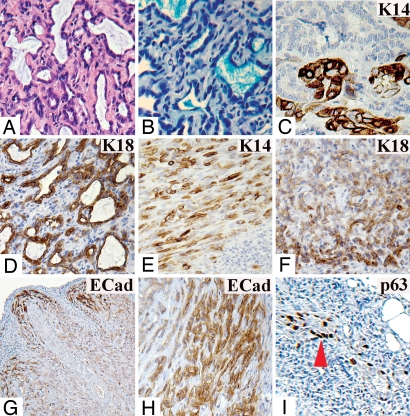
Fig. 2.
Staining of a tumor generated by LA7SL1. (A) H&E staining, core. (B) Alcian blue staining of tubule-secreted proteins. Antibody staining for: K14, core (C); K18, core (D); K14, periphery (E); K18, periphery (F); and E-Cadherin, panorama (G). (H) E-Cadherin antibody staining of a tumor generated by 100 sLA7 cells (periphery). (I) p63 antibody staining of tumor generated by 105 cells periphery (arrow, p63+ cells). (Magnifications: G, X5; F and I, X10; A, D, E, and H, X20; B and C, X40.)

