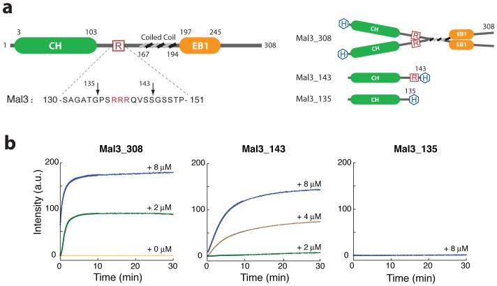
Figure 1.
Mal3 promotion of microtubule assembly in vitro. (a) Left, domain structure of Mal3. CH and EB1-like C-terminal motif domains are indicated by green and orange, respectively. Boundaries for each domain are indicated by residue numbers. The amino acid sequence for residues 130-150 is shown below and the three arginine residues are highlighted in red. Right, domain structure for the constructs used in this study. 6×His-tags are highlighted in blue. (b) Microtubule-polymerization assay by 90° light scattering at λ = 350 nm. All assays contained 4 μM S. pombe tubulin (heterodimer concentration). Colored lines indicate the added Mal3 monomer concentration; orange, 0 μM; green, 2 μM; brown, 4 μM; blue, 8 μM.

