Fig. 5.
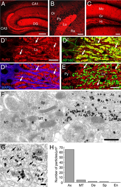
Preferential localization of the type 2 ryanodine receptor (RyR2) in non-terminal axons of hippocampal mossy fibers. (A–C) Immunofluorescence for the RyR2 in the hippocampus. (D) Triple immunofluorescence for the RyR2 (red), neurofilament-160 (NF160, green), and microtubule-associated protein-2 (MAP2, blue) in the stratum lucidum of the CA3 region. (E) Double immunofluorescence for the RyR2 (red) and type 1 vesicular glutamate transporter (VGluT1, green). Arrows in D and E indicate the intense RyR2 signal in putative axon bundles. (F and G) Silver-enhanced immunogold labeling for the RyR2. Arrows indicate the smooth ER in non-terminal axons. (H) The density of RyR2 immunogold labeling. Ax, axon; MT, mossy fiber terminal; De, dendrite; Sp. spine; En, endothelial cell; Scale bars: A, 200 μm; B and C, 50 μm; D and E, 10 μm; F, 500 nm; G, 100 nm.

