Figure 4.
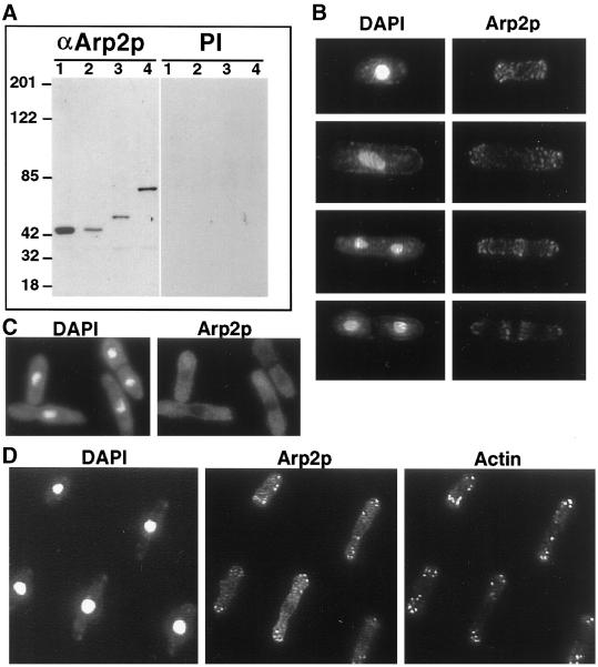
Immunolocalization of Arp2p. (A) Characterization of anti-Arp2p antibody. Lysates were prepared from wild-type (KGY28; lane 1), arp2-1 (KGY1187; lane 2), arp2-HA (KGY1524; lane 3), and arp2-myc (KGY1525; lane 4) strains and resolved by SDS-PAGE, then immunoblotted with anti-Arp2p or preimmune serum. (B) Wild-type cells (KGY28) were grown to midlog phase, fixed with methanol, and stained with a 1:5 dilution of affinity-purified rabbit polyclonal anti-Arp2p antibodies. S. pombe cells representing different stages of the cell cycle are shown stained with DAPI to visualize DNA and anti-Arp2p antibodies. Arp2p localizes to cortical actin patches at all stages of the cell cycle. (C) arp2-1 cells (KGY1187) were grown to midlog phase at 25°C and shifted to 36°C for 6 h. Cells were fixed with methanol and stained with affinity-purified anti-Arp2p antibodies as described above. Arp2p is diffusely distributed throughout the cytoplasm in the arp2-1 mutant. (D) Wild-type cells (KGY28) were grown at 25°C, fixed in methanol, and stained with a 1:5 dilution of affinity-purified rabbit polyclonal anti-Arp2p antibodies and a 1:50 dilution of monoclonal anti-actin antibodies.

