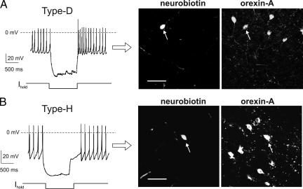
Fig. 4.
Two orexin cell types persist in different strains of mice. (A) (Left) Type-D fingerprint recorded in slice from wild-type (C57BL6J) mice. (Right) immunofluorescence imaging of the recorded cell, identified by neurobiotin staining; the cell contains orexin-A. Scale bar, 50 μm. (B) (Left) Type-H fingerprint recorded in slice from wild-type (C57BL6J) mice. (Right) immunofluorescence imaging of the recorded cell, identified by neurobiotin staining; the cell contains orexin-A. Scale bar, 50 μm.

