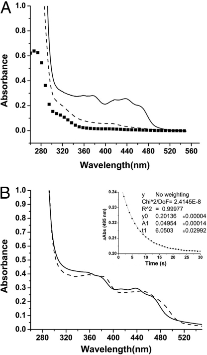
Fig. 1.
Absorption spectra and kinetics of copurified PixEHis-6–PixD. Solution containing both PixEHis-6 and PixD were bound to a nickel column and washed of unbound proteins under dark or light conditions. (A) Bound proteins were then eluted with imidazole under dark or light conditions and subjected to spectroscopic analysis. The solid line represents elution under dark conditions, and the dashed line is elution under light. The spectrum of PixEHis-6 alone is represented by squares. (B) Spectroscopic analysis of the dark-eluted PixEHis-6–PixD complex that exhibits a characteristic 10-nm light shift of the spectrum upon illumination. The solid line is a spectrum of dark-adapted protein, and the dashed spectrum is after white light illumination. (Inset) Decay kinetics of the PixEHis-6–PixD complex that exhibits a τ1/2 recovery to the ground state at ≈6 s, which is similar to the 5-s half-time of isolated PixD (11).

