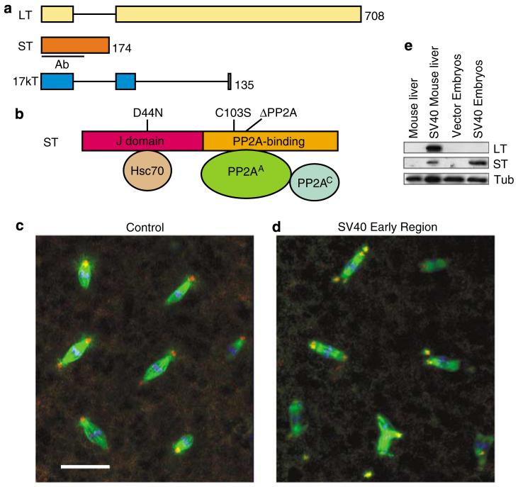
Figure 1.
Centrosome abnormalities in Drosophila embryos expressing Simian virus 40 (SV40) small tumor antigen (ST). (a) Schematic diagram of the SV40 early region gene products large tumor antigen (LT), ST and 17kT, produced by alternative splicing. Black lines indicate introns. Size of each protein (in amino acids) and region used to raise antibodies (Ab) are indicated. (b) ST has two domains: a J domain and a PP2A-binding domain. The mutations used in this study are indicated. (c, d) SV40 ST-expressing embryos exhibit supernumerary centrosomes and mitotic spindle abnormalities. Early cleavage stage embryos were stained for centrosomin (red), α-tubulin (green), and DNA (blue). Note some of the spindle poles are located outside of the image stack shown. Bar: 25 μm. (e) SV40 early region-expressing embryos produce ST protein, but no detectable LT. Lysate from mouse liver expressing SV40 early region (Comerford et al., 2003) is included as a positive control.

