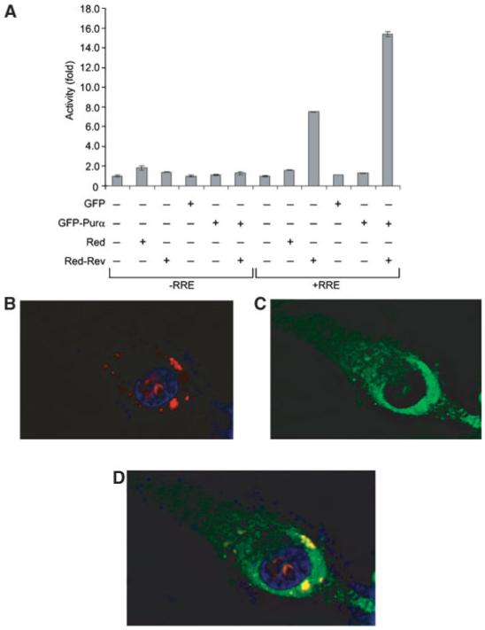
Fig. 3.

Subcellular localization of Rev and Purα in cells transfected with PLEGFP-Purα and pDs-Rev-Red1 plasmids. A: U-87MG cells were transfected with reporter vectors with or without RRE along with plasmids expressing fusion Red-Rev and GFP-Purα fusion fluorescent proteins in various combinations as denoted. Cells were fixed and red fluorescence from Rev (B) and green fluorescence from Purα (C) were detected by microscopy. Composite of the two colors demonstrates co-localization of Purα and Rev (D). DAPI Blue is for nuclear staining.
