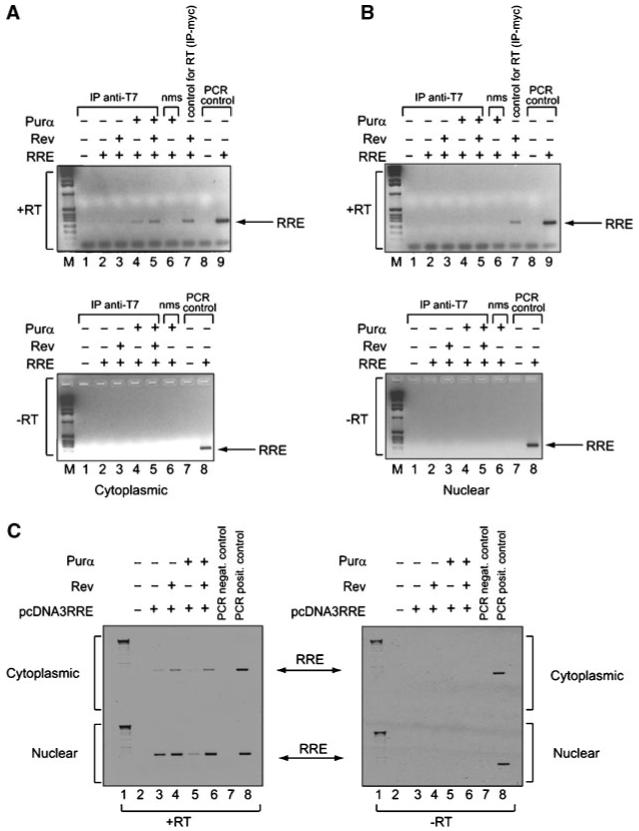
Fig. 8.

Interaction of Purα with RRE in vivo. Antibodies against Purα (anti-T7, lanes 1-5) and Rev (anti-myc, lane 7 in “+RT” panels) were used to immunoprecipitate interacting components from cytoplasmic (A) and nuclear (B) lysates from cells as described in Figure 7. Normal mouse serum (nms) was used as a negative control for immunoprecipitation (lane 6). RNA extracted from these complexes was subjected to RT-PCR with (“+RT”) and without (“-RT”) reverse transcriptase (RT) using RRE 1-234 specific primers. For PCR control, pcDNA3.1-SD4*-luciferase-RRE-SA7 and RRE 1-234 specific primers were used (lanes 8-9 in “+RT” panels and lanes 7 and 8 “-RT” panels). M: DNA marker. RNA input was verified by extraction of RNA directly from nuclear and cytoplasmic fractions (without prior immunoprecipitation) following RT-PCR (C).
