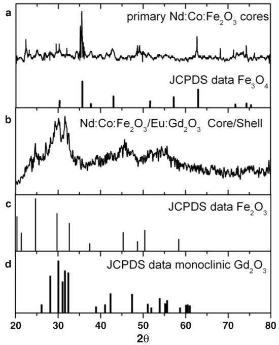
Figure 5.

(a) X-ray diffraction spectrum of the primary Nd:Co:Fe2O3 particles are compared to the typical XRD peaks of Fe3O4; (b) XRD of the core/shell Nd:Co:Fe2O3/Eu:Gd2O3 particles; (c) and (d) show the typical XRD spectral peaks of Fe2O3 and monoclinic Gd2O3 respectively.
